
What is the Spleen?
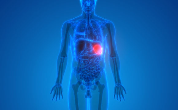
The spleen is a vital organ located in the upper left quadrant of the abdomen, typically weighing around 150 grams (0.33 pounds) in adults. It plays several crucial roles in the immune system and the management of blood cells. Conditions affecting the spleen, including splenomegaly and splenic rupture, may develop due to various causes and can affect its normal function. In severe cases, treatment may depend on the underlying condition and may require a surgical procedure such as spleen removal.
Functions of the Spleen
1. Immune Response:
– The spleen filters blood and removes old or damaged red blood cells and pathogens. It also produces lymphocytes, which help the body fight infections.
2. Blood Filtration:
– Acts as a reservoir for blood, where it can store red blood cells and release them into the circulation as needed—especially during stress or hemorrhage.
3. Iron Recycling:
– The spleen helps recycle iron from hemoglobin in old red blood cells, which is then used to produce new blood cells in the bone marrow.
Anatomy of the Spleen
-
Red Pulp:
- Composed of blood vessels and lymphatic tissue, responsible for filtering blood and facilitating the removal of old blood cells.
-
White Pulp:
- Contains lymphoid tissue that produces immune cells (lymphocytes) and mounts immune responses to infection.
Health Tips for a Healthy Spleen
To maintain optimal spleen health, consider the following lifestyle choices:
- Eat a Balanced Diet: Focus on fruits, vegetables, whole grains, and lean proteins.
- Stay Active: Regular physical activity supports overall immune function.
- Avoid Infections: Stay up to date with vaccinations and practice good hygiene to protect against infectious diseases.
Overview of Splenic Diseases
Introduction
Common Splenic Diseases
Splenomegaly
- Definition: Enlargement of the spleen due to various causes, including infections, liver disease, or blood disorders.
- Symptoms: Often asymptomatic but may include abdominal discomfort or pain in the upper left quadrant.
- Diagnosis: Physical examination, imaging studies (ultrasound, CT), and blood tests.
- Management: Treatment focuses on the underlying cause; splenectomy may be indicated if complications arise.
Hypersplenism
- Definition: A condition where an overactive spleen leads to excessive destruction of blood cells, resulting in reduced blood cell counts (anemia, thrombocytopenia, leukopenia).
- Symptoms: Symptoms often depend on the blood cell type affected; could include fatigue, easy bruising, and increased infections.
- Diagnosis: Blood tests showing low cell counts along with ultrasound or CT to examine spleen size.
- Management: Address underlying causes or consider splenectomy for symptomatic relief.
Splenic Abscess
- Definition: A localized collection of pus within the spleen due to infection.
- Symptoms: Fever, chills, left upper quadrant pain, and signs of sepsis.
- Diagnosis: Imaging studies (ultrasound, CT) to confirm the presence of abscesses.
- Management: Often requires antibiotics and, if large or symptomatic, surgical drainage or splenectomy.
Splenic Rupture
- Definition: A tear in the splenic tissue, often resulting from trauma or underlying disease (such as infections or tumors).
- Symptoms: Sudden onset of sharp pain in the left upper abdomen, hypotension, and signs of internal bleeding.
- Diagnosis: Immediate imaging (CT scan) and clinical assessment for signs of shock.
- Management: Emergency surgery (often splenectomy) is indicated to control bleeding and repair damage.
Splenic Tumors
- Definition: Tumors can be benign (e.g., hemangiomas) or malignant (e.g., lymphoma, splenic angiosarcoma).
- Symptoms: May present as splenomegaly or associated complications from the tumor.
- Diagnosis: Imaging studies and biopsies to ascertain histopathological type.
- Management: May involve surveillance, chemotherapy, or surgical resection depending on type and stage.
Symptoms to Monitor
Patients should be alerted to the following symptoms that may indicate splenic disorders necessitating further evaluation:
- Abdominal Pain Particularly in the left upper quadrant.
- Unexplained Weight Loss: Not linked to dietary changes or increased physical activity.
- Fever or Signs of Infection: Persistent fever or chills can indicate underlying issues.
- Easy Bruising or Bleeding : Signs that may result from blood cell deficiencies.
- Fatigue: Excessive tiredness not attributable to other causes.
When to Seek Medical Attention
Patients should consult a healthcare provider if they experience:
- Severe or persistent abdominal pain.
- Symptoms of splenic rupture, such as sudden sharp pain, hypotension, or signs of shock.
- Unexplained weight loss, fever, or changes in appetite.
Early diagnosis and intervention are crucial in managing splenic diseases and preventing complications. Patients with concerns about their splenic health should reach out to their healthcare providers for thorough evaluation and guidance.
Overview of Splenic Surgery
Introduction
Types of Splenic Surgery
- Definition: Splenectomy is the surgical removal of the spleen, which can be either partial or complete.
- Indications:
- Trauma: For ruptured spleen due to injury, which can lead to life-threatening internal bleeding.
- Splenic Tumors: For the excision of benign or malignant tumors requiring removal.
- Hypersplenism: When the spleen is overactive, leading to excessive destruction of blood cells.
- Spleen-related Complications: Such as abscess formation or splenomegaly that causes pain or discomfort.
- Surgical Technique:
- Overview: Can be performed via open surgery or laparoscopic techniques, with laparoscopic methods being less invasive and generally associated with quicker recovery.
- Procedure Steps:
- Anesthesia: General anesthesia is administered to the patient.
- Incision Types: A larger incision for open splenectomy; multiple small incisions for laparoscopic splenectomy.
- Resection: The surgeon identifies and removes the spleen while managing the connected blood vessels by ligating the splenic artery and vein.
- Closure: The remaining tissue is inspected for bleeding before closure.
- Duration: The laparoscopic procedure typically lasts about 1 to 3 hours, while open surgery may require additional time.
- Recovery:
- Hospital Stay: Patients usually remain hospitalized for 1 to 3 days post-surgery for observation.
- Postoperative Monitoring: Heightened surveillance for bleeding and infection, as well as assessment of overall recovery.
- Dietary Management: Normal eating patterns can be resumed once the patient is stable.
- Potential Complications:
- Common Risks: Include infection, bleeding, and complications related to anesthesia.
- Specific Complications:
- Infection: Increased risk of respiratory infections, particularly pneumonia, due to loss of splenic function; preoperative vaccinations may be recommended.
- Thromboembolic Events: Slightly elevated risk of developing blood clots postoperatively.
- Splenic Dysfunction: May impact the immune system for the lifetime of the patient, especially if large portions of splenic tissue are inadvertently removed.
- Definition: Splenic cyst deroofing is a surgical procedure aimed at treating symptomatic splenic cysts by excising the cyst wall (deroofing) while preserving the surrounding splenic tissue.
- Indications:
- Symptomatic Splenic Cysts: Patients who experience abdominal pain, fullness, or discomfort due to cyst size or pressure on adjacent structures.
- Complications from Cysts: Such as infection, rupture, or hemorrhage, necessitating intervention to prevent further complications.
- Diagnosis of Uncertain Cysts: For complex or atypical cysts that may require histological evaluation based on imaging findings.
- Large Cysts: Generally indicated for cysts exceeding 4 centimeters in diameter that are likely to cause symptoms or complications.
- Surgical Technique:
- Overview: Can be performed via traditional open surgery or minimally invasive laparoscopic techniques, depending on cyst characteristics.
- Procedure Steps:
- Anesthesia: General anesthesia is administered.
- Incision:
- Laparoscopic Approach: Multiple small incisions to insert trocars and utilize camera assistance.
- Open Surgery: A larger incision in the left upper quadrant, as necessary.
- Identifying the Cyst: The cyst is located under direct visualization with ultrasound or laparoscopic techniques.
- Deroofing: The cyst wall is excised, allowing its contents to drain while preserving splenic parenchyma, thus reducing the risk of recurrence.
- Closure: Laparoscopic incisions are closed with sutures or adhesive strips; in open surgery, the wound is similarly closed.
- Duration: The laparoscopic procedure typically lasts about 1 to 2 hours, while open surgery may take longer depending on complexity.
- Recovery:
- Hospital Stay: Patients usually remain hospitalized for 1 to 3 days for monitoring and recovery.
- Postoperative Monitoring: Careful assessment for signs of complications such as bleeding and infection.
- Dietary Management: Patients typically resume normal eating patterns shortly after surgery.
- Potential Complications:
- Common Risks: General risks include bleeding, infection, and anesthesia complications.
- Specific Complications:
- Infection: Risk of postoperative infection, especially in patients with pre-existing conditions or if the cyst was previously infected.
- Abscess Formation: May occur if the cyst is not adequately managed.
- Splenic Dysfunction: Minor risk of affecting splenic function, though substantial tissue removal is typically avoided.
- Recurrence of Cyst: There remains a risk of cyst recurrence; however, complete deroofing aims to minimize this.